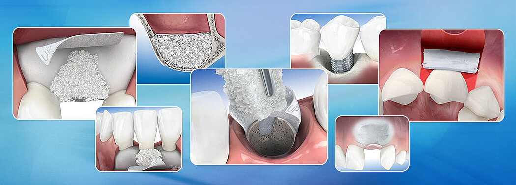
Treatments

Overview
At Geistlich Biomaterials, we are committed to developing treatments that are uniquely matched to the clinical situations you see every day. That’s why we do more than bring you a family of products – we provide proven solutions in specific therapeutic areas.
Advances in regenerative technology, biomaterials, and clinical techniques continue to create a growing number of treatment options for you and your patients. In order to consistently achieve successful long-term outcomes, it is critical to look beyond product characteristics and technical data. A true partnership means being empowered with scientific evidence empowerment, treatment planning tools, and dynamic educational resources.
Building this essential partnership with you, starts with information. Beyond the science that is fundamental to all of our biomaterials, we have also organized our family of products within distinct therapeutic areas.
References:
- Giannobile WV, et al.: Clin Oral Implants Res 2018; 29 Suppl 15: 7-10. (consensus report)
- Perrussolo J, et al.: Clin Oral Implants Res 2018 Oct 22 [Epub ahead of print]. (clinical study)
- Griffin TJ, et al.: J Periodontol 2006; 77: 2070-79. (clinical study)
- Soileau KM, et al.: J Periodontol 2006; 77: 1267-73. (clinical study)
- Zucchelli G, et al.: J Clin Periodontol 2010; 37: 728-38. (clinical study)
- Cairo F, et al.: J Clin Periodontol 2012; 39: 760-68. (clinical study)
- Sanz M, et al.: J Clin Periodontol 2009; 36(10): 868-76. (clinical study)
- Geistlich Mucograft® Seal Advisory Board Meeting Report, 2013. Data on file, Geistlich Pharma AG, Wolhusen, Switzerland.
- Thoma DS, et al.: J Clin Periodontol. 2020 Feb 24. doi: 10.1111/jcpe.13271. [Epub ahead of print]. (clinical study)
Biofunctionality: Based on Science
What Nature Inspires, Geistlich Engineers
Intentional design and the preservation of biologically natural structures are key elements in the development of Geistlich Biomaterials and the accompanying dental applications. Through unique, proprietary technology used during manufacturing, nature’s complex tissues are carefully processed to preserve biologic cues that enable optimal tissue integration.1,2
Biologically Natural Structures – Ideal Architecture
The crystalline structure of Geistlich Bio-Oss® and the Type I & III bi-layer collagen structures of Geistlich Bio-Gide® and Geistlich Mucograft® retain natural forms, allowing the body to accept these biomaterials as native. The surface of Geistlich Bio-Oss® supports the adsorption of proteins that enables adhesion of bone-forming cells.3-5 Using specific surface receptors, cells bind directly to Geistlich collagens. Geistlich Mucograft® is designed to provide a requisite, reinforcing matrix and a signaling source for regenerative wound healing. Fibroblasts respond to the collagen by attaching, orienting, and producing new collagen integration. Collagen research suggests that in such scaffolds, endothelial progenitor cells are activated for angiogenesis, and the intact collagen fibrils serve as conduits for endothelial cells and the formation of vascular channels of nutrition. These vascular channels are surrounded with perivascular mesenchymal stem cells with anti-inflammatory properties.6-9 Due to these properties, the clinical result observed with Geistlich Mucograft® is optimal soft tissue regeneration rather than soft tissue repair. 10-13
Nature’s Capacity for Healing
The human body possesses the capacity for regenerative wound healing. The natural structures of Geistlich Bio-Oss®, Geistlich Bio-Gide®, and Geistlich Mucograft® enhance the biologic cascade of healing events by attracting and delivering essential serum proteins. The result is complete tissue integration that encourages regenerative healing.
References:
- Rothamel D, et al., Clin Oral Implants Res. 2005; 16(3): 369-78
- Schwarz F, et al., Clin. Oral Implants Res. 2006; 17: 403-409
- Taguchi Y, et al., Biomaterials. 2005 Nov;26(31):6158-66
- Galindo-Moreno P, et al., Clin Oral Implants Res. 2014 Mar; 25(3):366-71. doi: 10.1111/clr. 12112 Epub 2013 Jan 28
- Araújo MG, et al., Clin Oral Implants Res. 2010 Jan; 21(1):55-64
- Nien Y, et al., Wound Repair and Regeneration. 2003; 11(5), 380-385
- Tran KT, et al., Wound Repair and Regeneration, 2004; 12(3), 262-268
- Davis GE, et al., Biochemical and Biophysical Research Communications. 1992 182(3), 1025-1031
- Tran KT, et al., Journal of Dermatological Science. 2005; 40(1), 11-20
- Badylak S, et al., 2009. Acta Biomaterialia, 5(1), 1-13
- Ghanaati S, et al. Biomed Mater. 2011 Feb; 6(1): 015010
- Rocchietta I, et al., Int J Periodontics Restorative Dent. 2012 Feb; 32(1):e34-40
- Nevins M, et al. Int J Periodontics Restorative Dent. 2011 Jul-Aug; 31(4):367-73
- Berglundh T, Lindhe J, Clin Oral Implants Res 1997; 8(2): 117–124.
- Becker J et al., Clin. Oral Implants Res. 2009; 20(7): 742–93
- Schwarz F et al., Clin Oral Implants Res. 2014 Sep;25(9):1010–5
- Weibrich G et al., Mund Kiefer Gesichtschirurg 4, 2000; 148–152
- Degidi M et al., Oral Dis. 2006 Sep; 12(5): 469–475
- Mordenfeld A et al., Clin Oral Implants Res. 2010 Sep;21(9):961–70
- Jung R et al., Clin Oral Implants Res. 2013 Oct;24(10):1065–73